- Rozwiązania dla Ciebie
- Obrazowanie
- Unity stomatologiczne
- CAD/CAM
-
Oprogramowanie
-
Planmeca Romexis
- Moduły oprogramowania
- Romexis 2D Imaging
- Romexis 3D Imaging
- Romexis 3D implantology
- Romexis Cephalometric Analysis
- Romexis 3D Cephalometry
- Romexis CMF Surgery
- Romexis CAD/CAM
- Romexis CAD/CAM Design
- Romexis Ortho Simulator
- Romexis Smile Design
- Romexis Dental PACS
- Bezpieczeństwo danych
- Charakterystyki
- Historie użytkowników
- Narzędzia AI dla stomatologii
- Wirtualna rzeczywistość
- Aplikacja mobilna obrazowania
- Darmowa przeglądarka obrazów
- Usługa przesyłania obrazów
- Aplikacja do przesyłania obrazów dla laboratoriów
- Usługa hostingu w chmurze
- Zasoby
- Wersje oprogramowania
- Do pobrania
- Szkoleniowe materiały wideo
-
Planmeca Romexis
- Opinie i doświadczenia użytkowników
- AI
- Dental Cabinetry
- Nowości & wydarzenia
- Szkolenia
- Bank materiałów
- veterinary
- Extranet
- Informacja Planmeca dotycząca prywatności
Obrazowanie 3D
Obrazowanie panoramiczne
Obrazowanie wewnątrzustne
Obrazowanie cefalometryczne
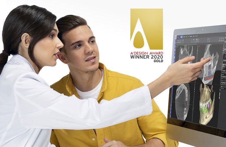
Planmeca Romexis® jest wszechstronną i intuicyjną platformą oprogramowania stomatologicznego typu all-in-one. Zapewnia ona dostęp do szerokiej gamy narzędzi spełniających zróżnicowane wymagania z zakresu obrazowania zarówno w przypadku małych klinik, jak i dużych szpitali.
Oprogramowanie do obrazowania stomatologicznego Planmeca Romexis®
Oprogramowanie Romexis typu „all-in-one” obsługuje najwięcej typów obrazowania 2D i 3D oraz CAD/CAM.
Aplikacje wspomagające
Posiadamy w swojej ofercie zróżnicowane i niezawodne oprogramowanie wspomagające wykonywanie, przeglądanie oraz przesyłanie obrazów.
Wydajne oprogramowanie dla klinik
Zwiększ wydajność pracy swojej kliniki dzięki aktualizowanym na bieżąco informacjom o zdarzeniach oraz danym dotyczącym użytkowania wszystkich połączonych w sieć urządzeń.