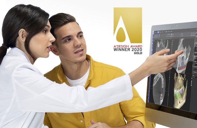
Romexis software release 6.4.5

We are excited to present the latest version of our software platform – Planmeca Romexis® 6.4.5. This release introduces new and powerful AI-based tools that not only help generate time savings for the clinician but also facilitate patient education and communication by visualising the cases better than ever before.

Highlights

menu-romexis-second-opinion.jpg

AI-based tool for 2D imaging: Second Opinion® integration

A seamless integration with the Second Opinion radiologic detection aid service allows Romexis users to receive AI detections created by the service directly in Romexis.

Find out more

menu-romexis-smart.jpg

AI-based tool for 3D imaging: Romexis® Smart

Romexis Smart enables the automatic segmentation and recognition of anatomies and the automatic mapping of CBCT images and intraoral scans.

Find out more

menu-romexis-new-3d-rendering-style.jpg

New 3D rendering style

A new type of rendering for CBCT images enhances the visualisation of cases.

Find out more

Second Opinion® integration

romexis-second-opinion-integration.jpg


Second Opinion® is a radiologic detection aid service provided by Pearl Inc. It is designed to aid dental health professionals to review the radiographs of permanent teeth in patients 12 years of age or older. Second Opinion helps to identify suspected dental findings which include:


  • Caries

  • Bone loss

  • Discrepancy at the margin of an

    existing restoration

  • Calculus

  • Periapical radiolucency

  • Widened periodontal ligament

  • Crown (metal, including zirconia & non-metal)

  • Filling (metal & non-metal)

  • Root canal

  • Bridge

  • Implants.


Second Opinion supports intraoral and panoramic X-ray images.


The Second Opinion service is cleared for clinical pathology detection for dentists in over 100 countries. For more information, please contact Pearl Inc.

The Planmeca Romexis® 6.4.5 release introduces an integration between Romexis and the Second Opinion service, which allows Romexis users to access the Second Opinion service seamlessly from Romexis and receive the AI detections created by the service directly in Romexis as image overlays.


Note: The service is provided, sold and invoiced directly by Pearl Inc. Before using the service, the doctor/clinic needs to contact Pearl Inc to create an account and a subscription to the Second Opinion service. For more information, please visit https://pages.hellopearl.com/romexis.


Disclaimer: Second Opinion utilises AI to generate proposals; however, it’s essential to emphasise that the ultimate decision-making authority remains with the doctor and the clinician.


There are two ways to use the service. Images can be sent either with a button click (on demand) or automatically whenever a new intraoral or panoramic image is captured, scanned, or imported to Romexis. Retrieving AI detections will take around 1–2 minutes. Regional servers are available for EU customers.

Features

Pathological and non-pathological findings

Second Opinion generates detailed labels around all potential pathologic and non-pathologic findings that it detects. The mouse hover will show the details of each finding. The user will evaluate the potential finding and delete any false positive findings if needed.

romexis-second-opinion-features-1.jpg


Bone level measurements

For intraoral images, the Second Opinion service provides the automatic measuring of bone level measurements.

romexis-second-opinion-features-2.jpg


Tooth parts

Second Opinion automatically segments and colours different parts of teeth (e.g. enamel, dentine, pulp, etc.) for easy visualisation and communication with patient for example to explain the advancement of caries into the dentine.

romexis-second-opinion-features-3.jpg


Tip!

Romexis magnifier is a great tool to zoom the labels and findings in demos and patient education. In Romexis 6.4.5 it now also zooms the annotations.

romexis-second-opinion-features-4.jpg


romexis-second-opinion-benefits.jpg

Benefits

Second pair of eyes

Dentists using Second Opinion gain a second set of eyes to help ensure that they have considered many common conditions that appear in their patients’ radiographs. That assurance comes from the system’s ability to detect signs of various dental pathologies and other treatable conditions found in dental radiographs – including hard-to-spot issues such as incipient caries or the early signs of a periapical radiolucency.

Second Opinion also operates as the doctor’s personal radiologists/assistant by annotating findings for easy review. Using Second Opinion boosts the efficiency, consistency, and accuracy of clinic’s radiographing to a new level.

Patient communication, retention and commitment

Findings and tooth anatomy are highlighted in text and colour establishing an easy-to-understand independent opinion that will be easy for the patient to relate to. This will help in gaining patient trust and will allow for maximising the clinic’s clinical performance by enabling the chairside detection of decay, infections, and numerous other conditions in patients’ X-rays. Ultimately, timely interventions can be performed, and long-term health can be provided to the patient base through increased patient retention.

Streamlined workflow with Romexis for unparalleled convenience

Through the seamless integration of Romexis and Second Opinion, users can effortlessly view AI annotations within the same Romexis software used for image capture, viewing, and storage. Users can easily send images with a single click or have all images automatically processed after capture and results in by the time finished capturing new images. As all images and findings are stored in Romexis, a permanent copy of all information is kept in one place.

Romexis® Smart

romexis-smart.jpg


Romexis® Smart is a new AI-based feature available for the Romexis 3D imaging module. It allows the automatic segmentation and recognition of anatomies, such as teeth, nerves, jaws, airways, and sinuses. Anatomies are cleanly segmented and coloured, which enables the easier and faster use of the software and provides an excellent visualisation of the case for patient education. With the help of artificial intelligence, CBCT images and intraoral scans are also automatically mapped. Thanks to the automatic tooth number recognition, a CBCT volume can be easily navigated just by clicking on the tooth number in the tooth chart and the software centres all views on the tooth of interest.


The AI feature of Romexis Smart has been approved for CE marking; other approvals are pending.


Disclaimer: Romexis Smart utilises AI to generate proposals; however, it’s essential to emphasise that the ultimate decision-making authority remains with the doctor and the clinician.

Features

Segmentations

Using Romexis Smart, anatomical structures such as teeth and airways and the maxilla, mandible, skull, soft tissue, and mandibular nerve can be automatically segmented into STL objects. The anatomies are visualised in the Romexis 3D rendering, and the visibility can be controlled in the object browser. All segmented objects can also be exported as STL models.

romexis-smart-features-1.jpg


CBCT and IOS mapping and fused tooth

Romexis Smart can automatically create a proposal for the intraoral scan and CBCT mapping. Furthermore, a more detailed segmented tooth can be generated by fusing together the crown from the intraoral scan with the root from the CBCT data.

romexis-smart-features-2.jpg


Tooth navigation

With the help of AI, Romexis Smart recognises the tooth numbers from the CBCT data. This enables the tooth number-based navigation in Romexis. Simply by clicking the tooth number in the tooth chart, all 2D views are centralised to that tooth in the Explorer and Implant/ Cross Section modules.

romexis-smart-features-3.jpg


Visualisation options for segmented anatomies

When the CBCT image is segmented, there are two options to visualise the segmented objects. The default option is the colourful one where all different anatomies are coloured with different colours. However, it is also possible to view the segmentations with more anatomic colours.

romexis-smart-features-4.jpg


romexis-smart-benefits.jpg

Benefits

Patient education

Segmented anatomies offer an excellent means of visualising anatomical structures and explaining treatment plans for patients. Demonstrating various aspects such as tooth extraction and implantation with visible roots and nerve-tooth connections, autotransplantation, sinus-tooth relations, diminished bone volumes, and airway structures may considerably boost case acceptance rates.

Implantology

The automated nerve detection and visualisation of adjacent teeth with roots assist in effective implant planning. The automatic fitting proposal for the CBCT and intraoral scan makes using the software easier and generates time savings.

Orthodontics

Romexis Smart enhances the benefits of using CBCT in diagnosing and treatment planning as root positions can be visualised in relation to the bone in a clear manner. This can help in more precise treatment planning and the avoidance of root collisions, dehiscence and fenestrations and materialise in fewer patient visits in total.

CMF Surgery

Romexis Smart substantially streamlines the pre-planning stages within the CMF surgery workflow, automating tasks such as jaw segmentation and CBCT integration with intraoral scans. This yields substantial time savings, enabling surgeons to focus on more engaging aspects, such as precise surgery planning. Additionally, a new feature in the Romexis® CMF Surgery module provides the visibility of segmented teeth and roots, offering invaluable insights into tooth root structures during the planning phase.

romexis-smart-benefits-cmf-surgery.jpg

New 3D rendering style

romexis-new-3d-rendering-style.jpg


romexis-rendering-options.jpg

Romexis 6.4.5 introduces a new type of rendering for CBCT images for even better visualisation than before. There will be three to four new presets.

Xfer

A new default rendering for new images.

romexis-xfer-3d-rendering-style.jpg


Transparent

The bone is more transparent and the soft tissues more visible.

romexis-transparent-3d-rendering-style.jpg


Endo

Designed for small volume sizes. The crowns are transparent and the dentine, root canal and pulp chamber more visible.

romexis-endo-3d-rendering-style.jpg


When a new rendering style is selected, there will be three new adjustments sliders:

• Adjust soft tissue opacity
• Adjust bone opacity
• Adjust teeth opacity

The brightness, contrast and threshold sliders are not visible when any of the new rendering styles are selected.

romexis-rendering-adjustments-slidersjpg.jpg